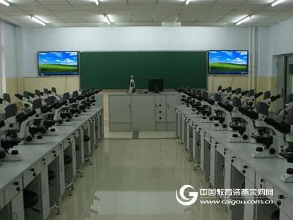
This article focuses on the features and benefits of the Motic Digital Microscopy Interactive Lab. Applied to pathology and histology teaching, it helps students to understand the structure of various tissues and organs, improve the coordination of students' brains and eyes, increase students' learning thinking ability, independent operation and analysis ability, and lay down for clinical learning in the future. Good foundation.
Key words: Motic digital; microscopy; interactive laboratory; basic medicine; experimental teaching
Experimental courses such as histology and pathology often use optical microscopes to complete experimental teaching under traditional laboratory conditions. It is difficult to achieve ideal goals for teacher-student interaction efficiency and teaching effects. Introducing the M otic digital micro-interaction laboratory in the teaching process of the medical morphology experiment course. While the students complete the self-handed observation of the microscopic specimens, they not only improve the teachers and students through digital camera, language interaction between teachers and students and multimedia network teaching. The efficiency of interaction, and the content of teaching has become richer, more innovative and more vivid.
1. The composition and characteristics of the laboratory
1.1 Composition
The teaching system is composed of image system, voice question answering system, digital microscope system and computer software system, including image processing and analysis module, multimedia teaching equipment, two-way voice communication system, digital microscope for students and digital multifunctional digital microscope for teachers. And software teaching platforms. The teacher and each student are connected to their respective computers through the U port to use a high-definition digital microscope, and the image processing units formed between them are relatively independent and powerful. LANs are interconnected between units. Equipment organization and classroom teaching are completed through a new distributed digital interactive software system. Image data sharing is comprehensive and voice communication is flexible.
1.2 Features
In terms of hardware, the students have their own computers, equipped with built-in integrated digital microscope, high effective pixels and resolution, and become an independent image processing platform; the high-end digital microscope with 5 million effective pixels can be given to the students. The courseware is played in real time; the hardware facilities formed are convenient to install and maintain, the network is stable and efficient, and the wiring is simple, which lays a foundation for better completion of the experimental class teaching.
In terms of software, the teacher can control each microscope and computer of the student, automatically turn off the computer, automatically open the application software of the student, monitor the student's computer screen in real time, and transmit the image of a student or teacher to all students. The students have discussed the teaching pointer to realize the dynamic real-time and the teacher to discuss the image under the microscope, communicate with the teacher through the text message mode, and the independent image processing analysis software package, effectively improve the students' independent analysis ability. The examination system reduces the labor intensity of teachers, realizes paperless examinations and automates the analysis of examinations.
In the teaching process of medical morphology experiment, through the Motic digital microscopic interactive teaching system, the teacher can observe the microscope picture of each student in the classroom in real time, and guide the students to correct the problems in the experiment through the voice question answering system. Students can output microscopic image and video signals to high-definition CCD cameras to output devices such as projectors and computers, which can visually display and discuss intuitive images with teachers and students. The teaching system has functions such as image processing, analysis, and digital video recording, which can save long-term preservation and reproduce relevant images and materials at any time, which is conducive to students' understanding of knowledge.
2. Characteristics and advantages of the laboratory
The Motic Digital Microscopic Interactive Lab teaching system has a clear picture and rich interactive means. Only one computer, through the local area network, can realize the interactive communication between teachers and students of images and voices, and solve the problems in the experiment process. By observing the student's microscope image in real time, the teacher can guide him to correct the problems in the real-time discovered experiment in time. Students can process microscopic and macroscopic images on their own computers in a timely manner. They can actively ask teachers for help through the questioning system. They can communicate with teachers and discuss with Motic's original microscope LED cursor arrows to indicate that the system is effectively and high-quality, and the communication between teachers and students is intuitive. Effective, it is conducive to the establishment of a good teacher-student relationship and the study and mastery of theoretical knowledge.
The Motic digital micro-interaction laboratory is equipped with a digital microscope for teachers. Students use digital microscopes. The two are connected by a distributor, data cable and corresponding software. The digital information displayed in the microscope field of the student can be displayed. On the computer hosted by the instructor, the teacher can project all the images onto the large screen through the projector. In this way, students not only see the images they observe through their own computers, but also see the images observed by teachers and other students, so that the digital image system can share the image resources between teachers and students.
The voice question answering system of Motic Digital Microscopic Interactive Lab is equipped with a set of earphones and call system for teachers and each classmate. During the teaching process, teachers and students can interact and communicate. All students can not only listen to the teacher's teaching and explanation, but teachers can also use the individual questions or group discussions to conduct experimental teaching according to the actual situation, and achieve a good teaching atmosphere of teacher-student interaction. The teaching model has also changed from “peer-to-peer†to “point-to-pointâ€, which has significantly improved teaching efficiency. Most students can think through a person's problem, thus mobilizing students' enthusiasm and initiative, exerting the main role of students and the leading role of teachers, reducing the labor intensity of teachers and improving work efficiency.
Motic Digital Microscopic Interactive Lab integrates specimen slides with large specimens to visually and accurately express abstract and complex teaching content through images, sounds and microscopes. Students receive the maximum acceptance in the shortest possible time. And to verify the theoretical knowledge of medicine, and to master it effectively, is conducive to the successful completion of teaching tasks.
The teachers and students of the Motic Digital Micro-Interaction Lab have the functions of static capture, automatic timing capture and dynamic video capture. Teachers can use this function to carry out the teaching activities of the experimental and difficult content. Students can take photos and record. Store important or difficult-to-understand microscopic images in your own computer and improve your scientific research capabilities.
3. Application in pathology and histology teaching
The use of the Motic digital microscopic interactive laboratory to establish a pathology and histology picture library and question bank can promote the smooth development of experimental teaching and better accomplish the objectives of the subject teaching.
In the teaching of pathology and histology, pictures are important teaching materials to help students master and identify various cells, tissues and organs. The traditional experimental teaching method has inconvenient access to the wall chart, which is easy to damage and lose. Teachers can establish a system image database stored in the chapters of teaching content by means of photography, scanning, etc. The missing structure in the slice can be replenished at any time during the experiment, and new tissue and pathological sections are continuously updated. It enriches the teaching content and can be used as a multimedia courseware for classroom teachers in classroom teaching.
Experimental assessment is a necessary means to test the quality of experimental teaching and reflect the ability of students. According to the question bank established by Motic Digital Microscopic Interactive Laboratory, the experiment can be carried out to comprehensively evaluate the students' learning effect.
The specific operation method of the assessment is:
(1) Generally, 5 minutes before the end of each experimental class, the teacher randomly selects 3 to 4 students to objectively evaluate the students' experimental ability and learning effect. Each time the unqualified students are assessed, the next lab will be retaken. Students will be organized for the final assessment before the final exam and the final exam results will be counted.
(2) Store test questions in the teacher's computer in advance, and conduct individual, group or whole class tests on a regular and irregular basis without prior notice, greatly improving the students' actual ability and level.
Beijing Khan Meng Zixing Instrument Co., Ltd.
June 22, 2017
Table Series,College Classroom Desk,Wooden Reading Table,Desks With Wheel
AU-PINY FURNITURE CO., LTD , https://www.aupinyjm.com